Digital approaches for beef carcass characterisation


Summary
- The European beef industry is increasingly seeking to adopt automation in meat processing.
- Recent advances in artificial intelligence (AI) offer significant opportunities to improve carcass characterisation, decision-making and automation within beef-processing environments.
- Meat Technology Ireland research at Teagasc is exploring the potential of sensors, imaging and AI for meat characterisation.
- Integration of multimodal sensing technologies including spectroscopy, 3D imaging, and CT scanning with AI can support the development of robust and intelligent digital meat characterisation and decision support.
The meat industry is increasingly seeking automation to address challenges such as labour shortages, worker welfare in abattoir environments, stringent hygiene requirements for non-contact processing and the need to minimise waste. Over the past few years, automation in poultry and pork processing has advanced considerably; however, the beef sector continues to lag due to several inherent challenges. Beef carcasses are substantially larger and exhibit significant biological variability, making the development of generic automated solutions particularly difficult. Consequently, many operations along the beef processing line remain either semi-automated or only partially automated. Tasks such as quartering, primal cutting, boning, and trimming still rely heavily on skilled manual labour. Manual processing introduces variability in cut quality, yield, and carcass classification accuracy, ultimately affecting profit margins and supply chain consistency. Meat Technology Ireland (MTI) is a major industry-led Technology Centre, hosted by Teagasc, that has a major pillar focused on meat digitalisation research. Within MTI, and internationally, current research efforts are increasingly focused on advancing automation through the integration of advanced computer vision systems and artificial intelligence techniques. Existing vision systems primarily rely on colour and depth information; however, they often lack the capability for detailed tissue-level carcass characterisation. Although X-ray-based techniques can provide insights into internal composition, their implementation requires expensive infrastructure, which presents a significant barrier for small- and medium-scale meat processors. In response to these challenges, research at Teagasc is focused on developing novel methodologies that integrate multimodal sensor data, including RGB imaging, depth sensing, and spectral information, to support decision-making systems for meat characterisation, decision support and future automation in beef processing. This article presents some of the recent developments in this area with a particular focus on meat characterisation.
Near-Infrared Spectroscopy (NIRS) for bovine tissue characterisation
Over the past three decades, NIRS has emerged as a valuable analytical tool due to its rapid acquisition speed, cost-effectiveness, and ability to analyse samples with minimal preparation. In meat science, NIRS has been extensively applied for evaluating chemical composition, predicting quality attributes such as tenderness and water-holding capacity, verifying meat authenticity, and detecting contaminants. More recently, machine learning (ML) algorithms have gained considerable attention in industrial automation, demonstrating superior capability over conventional chemometric methods in handling large, complex, and non-linear spectral datasets. As part of MTI, a study was conducted to investigate the potential of Visible–Near Infrared (Vis–NIR) spectroscopy in combination with several ML techniques to discriminate among major bovine tissue types. More than 300 samples of muscle, fat, marrow, bone, cartilage, tendon, ligament, and connective tissue were collected from a commercial beef-processing line. The spectra of the samples were pre-processed, and Principal Component Analysis (PCA) was conducted (Figure 1).
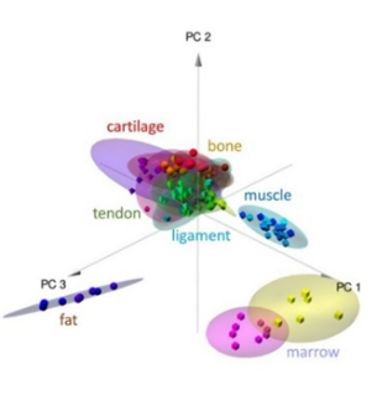
Figure 1. Tissue discrimination through PCA analysis
Results demonstrated clear separation of fat, marrow, and muscle tissue spectral signatures, whereas partial overlap was observed among ligament, tendon, cartilage and connective tissue samples due to similarities in their chemistry. Supervised classification models were developed using five machine learning algorithms: Random Forest (RF), Support Vector Machine (SVM), K-Nearest Neighbours (KNN), Linear Discriminant Analysis (LDA), and Partial Least Squares Discriminant Analysis (PLS-DA). These models were trained and tested using both the full wavelength range (FLW) and a reduced set of selected feature wavelengths (FW). Results demonstrated that LDA, PLS-DA, and SVM consistently outperformed RF and KNN models across most tissue categories. Comparable classification performance was achieved using both FLW and the selected FW, highlighting the feasibility of developing computationally efficient models for industrial implementation. Further analysis showed that models trained exclusively on NIR-range feature wavelengths achieved 100% overall classification accuracy across all evaluated classifiers, whereas models based solely on visible-range information exhibited reduced performance. These findings highlight the significant potential of NIR spectroscopy combined with ML techniques for accurate tissue-level carcass characterisation.
Carcass feature segmentation using deep learning
Recent advances in artificial intelligence (AI), particularly in deep learning, have created new opportunities for rapidly extracting and processing computer vision data to support robotic decision-making in beef processing environments. Convolutional neural networks (CNNs) have demonstrated significant potential for a wide range of object detection and segmentation tasks within meat-processing facilities. Within MTI digital pillar, the applicability of deep learning for beef carcass analysis, with pretrained models from two state-of-the-art frameworks (i.e. You Only Look Once (YOLO) and Detectron2), was tested using a diverse dataset comprising 118 carcasses collected from a commercial beef-processing plant. Images were acquired using two different imaging systems and developed models were trained to identify large anatomical structures (e.g. carcass, aitch bone, spine, and ribcage), and smaller and more complex features (e.g. individual ribs).
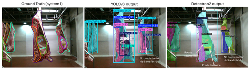
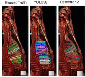
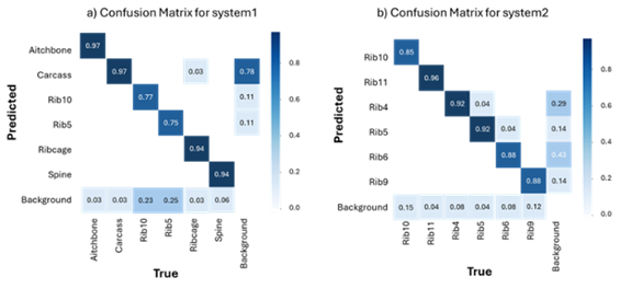
Figure 2. Prediction of carcass components through trained deep learning models
Results demonstrated that both frameworks achieved higher segmentation accuracy for larger anatomical structures compared with smaller features (Figure 2). The YOLOv8 model achieved mean Average Precision (mAP50; IoU = 0.5) values ranging from 94.1% to 97.0% for large structures, while rib segmentation accuracy ranged from 78.0% to 92.4%. Visual assessment indicated that smaller anatomical features were occasionally missed in background carcasses, contributing to lower mAP values, although segmentation performance improved significantly under controlled lighting conditions. Compared with Detectron2, the YOLOv8 framework produced sharper segmentation boundaries and demonstrated superior performance in identifying rib elements. Overall, these findings highlight the strong potential of pretrained deep learning models for automated beef carcass segmentation and characterisation.
Dimensional analysis through computed tomography (CT) and 3D imaging
Three-dimensional (3D) scanning and Computed Tomography (CT) are valuable technologies for obtaining precise dimensional and structural information from meat products. CT employs a rotating X-ray source to generate tomographic images based on tissue-specific attenuation coefficients, enabling the differentiation and quantification of tissues through reconstruction algorithms. As a result, CT is widely regarded as the gold standard for determining carcass lean meat content and is commonly used for calibrating other assessment instruments. Although both CT and 3D scanning are relatively time-consuming, they provide detailed information on carcass variability and can support the development of digital technologies for automated meat-processing applications. Short ribs, which are widely valued in international cuisines, contain varying proportions of lean, fat, connective tissue, and bone. Their dimensions are influenced by factors such as animal weight, category and carcass conformation. To support the development of automated processing systems for bone-in short rib products, a study was conducted using CT and 3D scanning data collected from steers, heifers, young bulls, and cows. Short rib sections corresponding to the 1st to 5th ribs were collected from 42 carcass sides obtained from a commercial beef-processing plant.
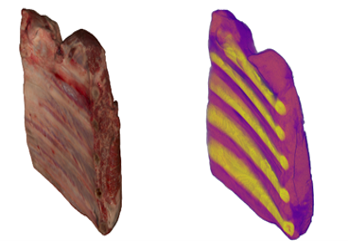
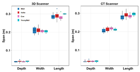
Figure 3. 3D scan, 3D constructed Computed Tomography (CT) scan, and Comparison of dimensions between both modality
Three-dimensional images were acquired using a Canon Aquilion Lightning X-Ray CT scanner and an Artec Leo 3D scanner. The surface meshes generated from both technologies provided information on short rib dimensions and rib bone positions. The dimensional distributions across animal categories were generally similar, although short ribs from young bulls were slightly longer compared to other categories. Measurements of short rib depth, width, and length obtained from both CT and 3D scanning showed strong agreement, with no significant differences observed at the 95% confidence level. Bland–Altman analysis further demonstrated that measurement differences were randomly distributed within acceptable limits. Slightly greater variation was observed for width measurements compared with depth and length. Overall, the results indicate strong agreement between CT- and 3D-derived measurements, suggesting that data from either system can be used to generate accurate digital representations of short rib products. Such information is valuable for understanding biological variability and product deformation behaviour, supporting the development of automated meat-processing systems.
Further reading
Mishra, J.P., Ferragina, A., Hegarty, S. and Hamill, R.M. (2026). Advancing bovine tissue discrimination with Vis–NIR spectroscopy coupled with machine learning methods. Food Production Processing and Nutrition 8: 27 (2026). https://doi.org/10.1186/s43014-026-00382-z
Mishra, J.P., Ferragina, A., Hegarty, S. and Hamill, R.M. (2026). Instance segmentation of beef carcass features with deep learning. Applied Food Research 6: Issue 1, 102070. https://doi.org/10.1016/j.afres.2026.102070
Acknowledgements
The authors thank Georgios Anagnostou and the staff of Liffey Meats and acknowledge the contribution of Meat Technology Ireland (MTI), a co-funded industry/Enterprise Ireland Technology Centre hosted by Teagasc (contract TC-2021-0031) and a Walsh Scholarship to Jyoti Mishra (WS2021039).
Compiled and edited by Mark McGee and Paul Crosson, Teagasc, Grange Animal & Grassland Research and Innovation Centre, and first published in BEEF2026 – Driving Sustainable Performance, additional reading from BEEF2026 is available here.